
News
Low folate levels in pregnant women may be linked to hyperactive children
30 April 2010
Attention-deficit and hyperactivity problems in children may be associated with low vitamin B9 (folate) levels during pregnancy, suggests a new UK study.
01 January 2014
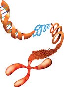
Cell aging is a complex process in which numerous factors can be involved. Telomeres located at the ends of chromosomes play an important role here – they consist of repetitive DNA sequences and associated proteins (histones) that stabilize the DNA by forming a kind of protective cap. When DNA is copied during cell division, some of the DNA building blocks (nucleotides) at the ends of the chromosomes are not copied, with the result that the telomeres on the newly formed DNA strands get shorter with each cell division. With increasing telomere shortening the cell changes its pattern of gene activation, slows its rate of division, then halts division completely (senescence), and eventually dies (apoptosis). Telomerase, an enz- yme that forms telomeres, counteracts this cell aging process by adding nucleotides that would otherwise be lost to the ends of the new DNA strand. Cell aging and death are thus delayed. Both telomere length and the amount and activity of telomerase, along with other factors, determine how many times cells can divide. Studies have shown that these factors can be positively influenced by micronutrients.
 Detectable amounts of telomerase are only found in the human body in cells that need to be constantly renewed, such as precursor cells (stem cells) and germ cells (gametes), as well as in tumor cells. While the latter have short telomeres, they pos- sess large amounts of telomerase. This, together with the particular genetics of tumor cells, ensures that they do not age naturally and can continue to divide. Research is now aiming to encourage telomerase synthesis or activity in healthy body cells in order to delay aging. It is also looking for ways to switch off telomerase activity in tumor cells (where present), in order to counteract the formation of new tumor cells. Micronutrients could play a role in the regulation of telomerase. Furthermore, antioxidant micronutrients could help to protect telomeres from damage caused by oxidative stress and thus contribute to a cell’s life span.
Protection of telomeres by antioxidants
With each cell division, and thus with every duplication of the genetic information carrier DNA, the chromo- somes lose pieces of functional units (telomeres) located at their ends. Telomeres appear to increase the stability of the genetic information and seem to be essential for the cell’s ability to divide (replication). The shortening of the telomere length that occurs with each cell division results in a change in the DNA, in its associated proteins ( histones), and in gene expression in the vicinity of the telomeres. When the ends of chromosomes reach a critical length, the cell loses its ability to replicate and dies (apoptosis). Numerous findings from in vitro studies speak for the hypothesis that the length of telomeres is associated with the aging process: the faster telomere length decreases, the faster a cell dies (1). At the same time, the risk of genetic changes and the emergence of life-shortening diseases seem to increase with the gradual loss of telomeres (2–5). Aside from the number of biologically limited cell divisions, telomere length may also be influenced by oxidative stress. Oxidative damage to proteins that are associated with telomeres and which interact with DNA repair enzymes (6), for example, may contribute to a rapid reduction in the length of the chromosome ends (7). Telomeres also seem to be particularly sensitive to free radicals in comparison with other DNA segments (8). Free radical formation is especially promoted by inflammation (9), as well as by environmental factors such as UV radiation, ozone, particulate matter, and tobacco smoke. Consequently, micronutrients with antioxidant properties that counteract the formation of free radicals or neutralize any free radicals formed could protect telomeres from oxidative damage and premature shortening.
The analysis of data from a cohort study with pairs of sisters showed that a higher intake of vitamin C and E and beta-carotene from food and nutritional supplements among the participants was accompanied by longer telomeres in leukocytes (white blood cells) compared with women with a worse supply of these vita- mins (10). Moreover, an increased intake of vitamin B12, which causes an increased availability of the antioxidant glutathione, was linked with comparatively long telomeres. A case-control study of over 200 wo- men reported that particular subjects with inadequate supply of antioxidant micronutrients after the meno- pause presented shortened telomeres and an increased risk of breast cancer (11).
The antioxidant and anti-inflammatory properties of the omega 3 fatty acids docosahexaenoic acid (DHA) and eicosapentaenoic acid (EPA) could counter the shortening of telomeres in cells involved in the defense against inflammations (12). A cohort study with over 600 participants showed that high plasma levels of DHA and EPA were linked to less incidence of telomere shortening (13). In a randomized controlled trial among healthy overweight men who took very little daily exercise, an increase in omega 3 fatty acid intake com- bined with a reduction in omega 6 fatty acid consumption led to telomere lengthening (14). A randomized controlled study of patients with mild cognitive impairment suggested that targeted doses of DHA could lead to an increased DHA concentration in red blood cells and a decrease of telomere shortening in cells (15).
For magnesium and zinc, in vitro studies and animal experiments provided evidence that these could po- tentially protect against telomere damage by reducing oxidative stress and inflammation (12, 16). In a co- hort study, longer telomeres were observed in women with a good supply of magnesium (10).
Protection of telomeres by vitamin D and B vitamins
Oxidative stress and inflammation are key determinants in the biology of aging and appear to be closely linked with telomeres. Thus, the anti-inflammatory properties of vitamin D may also contribute toward protection against telomeric shortening in lymphocytes (white blood cells) (17). In a large cohort study using twins, the vitamin D levels in the blood and the telomere length in leukocytes were measured (18) in a sub- set of the total of 2,160 women. It was found that higher vitamin D serum levels were associated with longer telomeres. Short telomeres are considered to be a risk factor for cardiovascular disease (19). Vitamin D’s hypothesized capacity to prevent cardiovascular disease, which has been demonstrated in studies, may be explained by the stabilizing effect of the vitamin on telomeres: vitamin D reduces proinflammatory mediators (e.g., TNF alpha and interleukin 2), which appear to have a detrimental effect on telomeres (18). In addition, a case-control study of patients with the autoimmune disease Lupus erythematosus showed a correlation between vitamin D deficiency and reduced telomere length on the one hand, and a subsequent extension of telomeres with improved vitamin D status on the other (20). A case-control study with hemodialysis patients had previously delivered similar indications (21).
Results of in vitro studies have provided evidence that vitamin B3 (nicotinamide) could affect the length of telomeres through a variety of mechanisms relevant to DNA stability. For example, the vitamin seems to effect the formation of DNA repair enzymes (22–25). Folate ( vitamin B9) could play a special role in the formation and maintenance of telomeres. The vitamin forms precursors of the DNA building blocks (nuc- leotides) and affects the stability of the DNA, of associated proteins, and of the telomeres. An inadequate supply of folate in the diet could lead to a shortening of telomeres, most likely due to increased DNA damage (15, 26). Folate deficiency may also cause an imbalance in available nucleotides during cell division and thus lead to a shortening of the telomeres due to incorrect DNA replication (27). Studies have shown that due to folic acid deficiency, the transfer of methyl groups to specific nucleotides and associated proteins (methy- lation) is reduced and this could impair the regulation of gene activity that controls telomere length, among other things (28). Over-long telomeres may sometimes result in cells where the activity of the telomere-lengthening enzyme (telomerase) is particularly high (e.g., in stem cells, germ cells, T lymphocytes, and certain tumor cells) (18, 26, 29). Cells with excessively elongated telomeres and reduced methylation-influenced regulation seem to be prone to unwanted reconnection of genetic material (recombination) (30). In addition, increased plasma levels of the amino acid homocysteine may occur due to insufficient folate levels in the blood (31). An increase in homocysteine levels has been associated with telomere shortening (26). An exception to this is seen at very high homocysteine concentrations, under which extended telomeres were observed. This can presumably be attributed to reduced DNA methylation and thus limited telomere regulation.
Influencing telomerase activity
The enzyme telomerase regulates the formation and length of the telomeres at the ends of chromosomes. These are essential for the stability of the DNA, particularly during the copying of genetic information that occurs when cells divide. Telomerases not only promote the lengthening of telomeres in germ cells and stem cells, but also (in certain cells) the repair of damaged sections of DNA. A telomerase deficiency may, there- fore, also result in increased chromosomal instability and the degeneration of cells (into cancer cells) (32– 36). On the other hand, an excessively high activity of the enzyme can confer upon tumor cells the ability to divide indefinitely. In such cells, naturally pre-programmed cell death (apoptosis) is switched off, meaning that they can multiply arbitrarily. Medical science is attempting to counteract cell division in telomerase- dependent tumors using telomerase inhibitors (37).
Since the ends of chromosomes become shorter with each cell division and the life of the cell is thus redu- ced, protecting telomeres from ( oxidative) damage and increasing telomere-lengthening telomerase activity are both important factors for delaying the cellular aging process. Studies have shown that telomerase activity can be significantly increased through simple changes in daily habits (38). It seems that mental stress is of particular significance here: reducing or normalizing stress (e.g., with relaxation exercises) may increase telomerase activity significantly. Telomerase activity can also be increased by reducing LDL cholesterol levels, normalizing the diabetic metabolism, and through weight loss if overweight.
Research into which micronutrients may most clearly positively affect telomerase activity is still in its infancy. According to the results of in vitro studies, the antioxidant vitamins E and C appear to stimulate telomerase activity (39, 40). The same might also be true for folate ( vitamin B9) (26, 28). Studies of the effects of dietary changes on enzyme activity in healthy subjects took into account, among other things, targeted doses of vitamin E (100 IU/day), vitamin C (2 g/day), as well as omega-3 fatty acids (3 g of fish oil/day) and selenium (200 µg/day) (41). Likewise, significantly increased telomerase activity in immune cells was observed in patients with a slightly aggressive form of prostate cancer who over five years had changed their lifestyle and had, for example, adopted a micronutrient-rich diet that included plenty of fruits and vegetables (42).
In a randomized controlled trial with severely overweight (obese) men and women, participants were given 2000 IU vitamin D or a placebo daily for four months (43). In the vitamin D group, the telomerase activity in peripheral mononuclear blood cells was significantly increased compared to the placebo group. Additional studies of patients with ovarian cancer suggest that targeted doses of vitamin D could down-regulate telo- merase activity in cancer cells and induce apoptosis (44). It has also been proposed that magnesium (16) and zinc might be involved in the regulation of telomerase activity. The ability of zinc to modulate telo- merase has so far been tested primarily in tumor cells (45, 46). The results suggest that the trace element could possibly both activate and deactivate telomerase.
30 April 2010
Attention-deficit and hyperactivity problems in children may be associated with low vitamin B9 (folate) levels during pregnancy, suggests a new UK study.
26 July 2009
The American Academy of Dermatology (AAD) is backing efforts to encourage increased consumption of vitamin D through food and dietary supplements.
3 November 2014
According to a new study from Canada children who drink non-cow’s milk such as rice, almond, soy or goat’s milk, are twice as likely to have low blood levels of vitamin D.